
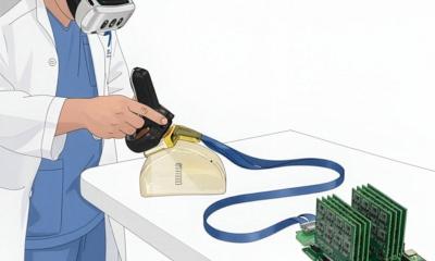
Every person carried out identification duties utilizing 4 totally different ultrasound applied sciences. In a single situation, they seen 2D photos on a daily display screen, which is the best way that almost all ultrasounds are actually carried out. In addition they seen 3D photos on a daily display screen, in addition to two augmented actuality circumstances: one 2D and one 3D (AR-VIU).
In a single spherical of experiments, customers had been requested to establish an object embedded in gelatin — reminiscent of a spring, a ball, or a screw — inside an opaque container that was scanned with ultrasound. In a second set, they had been requested to make use of a pen to mark the placement of “tissue phantom” — a gel-like materials engineered to imitate human tissue. This simulates the duty of finding the fitting spot for a needle throughout a biopsy.
The researchers discovered that the AR-VIU system considerably improved all customers’ capacity to establish and find objects. The impact was particularly sturdy for novices, who carried out practically in addition to consultants when utilizing AR-VIU. When utilizing the standard 2D imaging system, consultants carried out significantly better than novices. “Overlaying photos with the anatomy and offering 3D visible context makes ultrasound considerably simpler for novices to grasp,” Viswanath says.
In interviews after the experiments, many of the novices reported that they most well-liked the AR-VIU method, with many saying that it made the duties simpler. “The 3D system imposes much less mind drain, it’s extra intuitive, and it’s simpler to grasp what is occurring within the focused area,” Dagdeviren says.
Most of the consultants stated they most well-liked the standard 2D imaging as a result of that’s what they had been accustomed to and had been educated to make use of. Nevertheless, these consultants additionally stated they may see the advantages of the AR-VIU system in some conditions, reminiscent of putting a needle for a biopsy or visualizing the motion of the center wall throughout echocardiography.
The researchers are actually engaged on additional enhancing the decision of the imaging and doing further exams to show the accuracy of the AR-VIU know-how.
Supply: Massachusetts Institute of Expertise